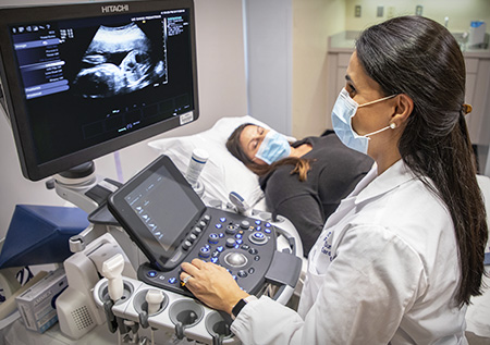
Fetal Echocardiography: A Dedicated Assessment of the Fetal Heart
Congenital heart disease is the most common category of structural birth defect, affecting approximately eight in every thousand live births. Many of these conditions can be identified before birth through a dedicated examination known as fetal echocardiography — a specialised ultrasound of the fetal heart that goes considerably beyond what is assessed during a routine anomaly scan. At Fetocare, fetal echocardiography is performed by Dr. Shubham Gupta with the precision, patience, and clinical depth that this examination demands. It is a scan that deserves unhurried time — and that is exactly what we give it.
What Is Fetal Echocardiography?
Fetal echocardiography is a targeted ultrasound examination that produces detailed, real-time images of the developing heart and its associated vessels. Unlike the cardiac views included in a standard anomaly scan — which provide a general overview of heart structure — a dedicated fetal echo involves a systematic, comprehensive assessment of every chamber, valve, vessel, and flow pathway in the fetal cardiac system. It uses both two-dimensional imaging and Doppler techniques to evaluate not only anatomy but also function, rhythm, and haemodynamics. The examination is carried out transabdominally, is entirely safe, and involves no radiation.
When Is It Recommended and What Does It Assess?
Fetal echocardiography is most commonly performed between twenty-two and twenty-six weeks of gestation, when the heart is large enough to be assessed in detail but still in a phase of development where findings can influence clinical planning. It is recommended in a number of situations, including:
- An abnormal or suspected abnormal cardiac view on the routine anomaly scan
- An elevated NT measurement at the first-trimester scan
- A family history of congenital heart disease (in a parent or sibling)
- Maternal conditions such as pre-gestational diabetes, lupus, or exposure to certain medications known to increase cardiac risk
- A chromosomal abnormality identified through prenatal testing, many of which carry an associated cardiac defect
- Twin-to-twin transfusion syndrome or other complex multiple pregnancy conditions
During the examination, all four cardiac chambers, the inflow and outflow tracts, the great vessels (aorta and pulmonary artery), the heart valves, and the septal walls are assessed systematically. Fetal cardiac rhythm and rate are evaluated, and Doppler assessment of flow across valves and through major vessels provides information about functional adequacy alongside structural findings.
What to Expect and Why It Matters
The examination typically takes between forty-five and sixty minutes, reflecting the thoroughness required. You will be asked to lie comfortably while the probe is moved gently across the abdomen. The baby's position and cooperation are important factors — if the heart is not in an optimal position for imaging, our team will wait and try again rather than compromise the quality of the assessment. After the scan, findings are discussed with you in full, with any abnormalities explained clearly, including their significance, the range of possibilities, and the next steps that would be appropriate.
Prenatal diagnosis of congenital heart disease makes a meaningful difference to outcomes. It allows parents and medical teams to plan delivery in an appropriate setting, ensures that paediatric cardiologists and surgical teams are informed and ready, and gives families the time to understand what lies ahead. At Fetocare, we approach every fetal echocardiogram with the knowledge that the information it provides can change the trajectory of a child's care — and we treat it accordingly.