3D / 4D Ultrasound: Lifelike Imaging With Clinical Purpose
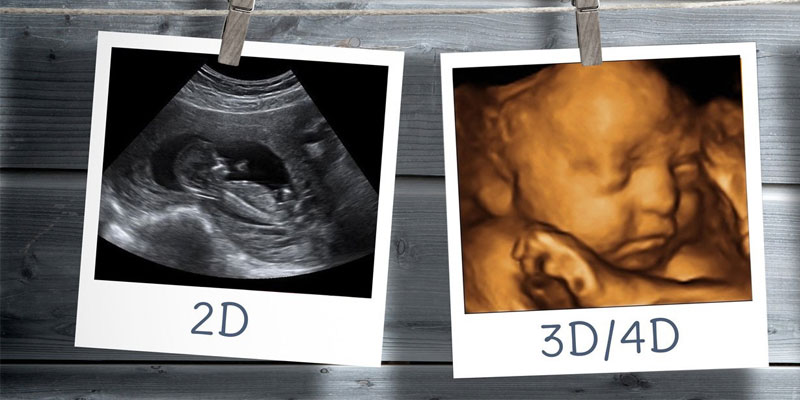
Three-dimensional and four-dimensional ultrasound represent a remarkable evolution in prenatal imaging. Where conventional two-dimensional scans provide flat, cross-sectional views of the baby, 3D ultrasound produces detailed, three-dimensional surface images, and 4D imaging adds the dimension of time — showing the baby in real motion, yawning, swallowing, and moving its limbs. At Fetocare, we offer 3D and 4D imaging not as a novelty, but as a clinically meaningful extension of our diagnostic capabilities — offered thoughtfully, where it adds genuine value to the care we provide.
What Is 3D / 4D Ultrasound?
Standard ultrasound — the type used throughout most prenatal scanning — produces two-dimensional images by sending sound waves through the body and reading the echoes that return. 3D ultrasound builds on this by acquiring multiple 2D images in rapid sequence from different angles and combining them into a three-dimensional rendering of the baby's surface. 4D ultrasound does the same in real time, at a speed sufficient to capture movement fluidly. The result is an image that looks far more like a photograph or video of the baby than the cross-sectional slices that parents have historically seen on a monitor. Both modalities use the same ultrasound technology and carry no additional risk to mother or baby beyond that of conventional scanning.
When Is It Performed and What Does It Show?
3D and 4D imaging is most effective between twenty-four and thirty-two weeks of pregnancy, when there is typically a good balance between fetal size and the amount of amniotic fluid surrounding the baby. Earlier than this, the structures are small and detailed surface imaging is limited; later, the reduced fluid and the baby's position can make clear imaging more difficult. Within this optimal window, 3D and 4D ultrasound can reveal:
- Facial features — including the nose, lips, eyes, and chin in remarkable detail, which can aid in the detection of cleft lip or other facial abnormalities that may not be clearly visible on 2D imaging
- Fetal behaviour and movement — swallowing, stretching, facial expressions, and limb movement can all be seen in real time on 4D imaging
- Surface anatomy — the spine, limbs, and external structures can be viewed from multiple angles, which may supplement the 2D anomaly scan findings
- Bonding and reassurance — for many parents, seeing their baby's face in three dimensions provides a profound emotional connection that supports the experience of pregnancy
What to Expect During the Scan
A 3D / 4D scan is conducted transabdominally, in the same manner as a standard ultrasound. You will be asked to lie comfortably while a handheld probe is moved gently across the abdomen. The quality of 3D and 4D images is significantly influenced by the baby's position at the time of scanning — if the baby's face is turned toward the placenta or is low in the pelvis, obtaining clear surface images may be challenging. Our team will take the time necessary to work around the baby's position, and will explain honestly what can and cannot be seen during the appointment. The session typically takes between thirty and forty-five minutes.
Our Approach at Fetocare
We believe that 3D and 4D imaging is most valuable when it is performed with clinical intent rather than as a standalone keepsake experience. At Fetocare, the 3D / 4D scan is always conducted alongside a thorough 2D assessment of fetal wellbeing, ensuring that the appointment serves both the emotional experience of the parents and the clinical oversight of the pregnancy. Every session is reviewed by Dr. Shubham Gupta, and any noteworthy findings are discussed with you directly. You leave not only with beautiful images of your baby, but with a full clinical update on how the pregnancy is progressing.